
Understanding Fundal Height in Pregnancy: Definition and Importance

Written by Arpan Ghosh
Reviewed by Dr. Thongram Biju Singh, MD


Key Takeaways
- Introduction
- How to Measure Fundal Height: Step-by-Step Guide
- Fundal Height by Week Chart: Normal Ranges and What to Expect
- Placenta Fundal Meaning: Understanding Placenta Position
- Placenta Position Monitoring: Why Placenta Location?
- Fundal Palpation in Pregnancy: Technique and Clinical Use
- Fundal Height After Delivery: What Changes to Expect
- Fundal Height Calculator Tools: Using Technology for Pregnancy Monitoring
- FAQs

Introduction
Have you heard of doctors using a tape to measure a pregnant woman's belly? This is a common phenomenon that has always been taken lightly but offers valuable information about the progress of pregnancy and foetal well-being. That size is referred to as fundal height and serves a significant role in monitoring the development of the baby and the uterus.
The height of the uterus, also referred to as the fundal height, is a necessity in the pregnancy monitoring process. It assists medical professionals in monitoring foetal development, identifying possible complications at an early stage, and ensuring the health of the mother and child. Understand more about fundal height in pregnancy with this blog.
How to Measure Fundal Height: Step-by-Step Guide
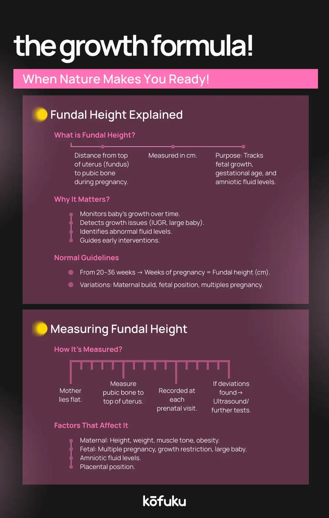
Fundal height measurement is a simple practice performed in prenatal clinics. Here’s how it's done:
- The mother lies flat on her back with her legs slightly bent.
- The healthcare provider locates the top of the pubic bone.
- Using the palm, they identify the fundus, which is the top of the uterus.
- A measuring tape is placed from the pubic bone to the top of the uterus.
- The number in centimetres represents the fundal height, usually corresponding to gestational weeks.
Knowing the correct way to measure fundal height helps achieve effectiveness. For example, at 24 weeks, the uterus is usually 24 cm in size. The deviations can reflect a condition such as intrauterine growth restriction or overall amniotic fluid.
This method, often called fundal palpation in pregnancy, is a non-invasive, cost-effective, and widely used clinical tool in obstetrics.
Fundal Height by Week Chart: Normal Ranges and What to Expect
Doctors often refer to a fundal height by week chart to compare actual measurements with expected norms.
- 12 weeks: uterus reaches just above the pubic bone.
- 20 weeks: the uterus aligns with the belly button.
- 28 weeks: The uterus lies midway between the navel and the rib cage.
- 36 weeks: the uterus reaches near the rib cage.
- 40 weeks: The uterus may descend slightly as the baby engages for delivery.
A reliable fundal height chart helps determine whether the foetus is developing normally. Minor differences are usual, but when they are quite considerable, additional scans are necessary.

Placenta Fundal Meaning: Understanding Placenta Position
The term fundal placenta appears in the reports of many women during ultrasound scanning, and they are curious to know whether it has any relevance. A placenta fundal indicates that the placenta has taken position against the upper part of the uterus, where the upper edge is the fundus.
This location is known for being safe and spacious, providing the baby with ample room to grow. The term placenta fundal just indicates the location of the placenta and not a medical problem.
Placenta Position Monitoring: Why Placenta Location?
The placenta plays an essential role in supplying the foetus with nourishment and oxygen. It is therefore vital to monitor its location. When reports state the placenta position as fundal, it means the placenta lies in the upper uterus. This is generally favourable compared to low-lying placentas, which may complicate delivery.
Fundal Palpation in Pregnancy: Technique and Clinical Use
Fundal palpation in pregnancy is a method of testing during which the medical professional uses their hands to examine the upper part of the uterus. It assists in the determination of
- The size and growth of the uterus.
- The position of the foetus.
- Whether the foetus has engaged before labour.
This physical examination, in combination with scans, provides a complete health overview of the mother and foetus.

Fundal Height After Delivery: What Changes to Expect
After childbirth, the uterus gradually shrinks back to its pre-pregnancy size, and the fundal height after delivery decreases daily.
- On day one after delivery, the uterus is usually at the navel level.
- By the end of the first week, it drops into the pelvic cavity.
- Within six weeks, it typically returns to normal size.
Observation of the postpartum height of the uterus is necessary to rule out complications like retained products or uterine infection.
Fundal Height Calculator Tools: Using Technology for Pregnancy Monitoring
The fundal height calculator can be of benefit to both mothers and doctors with the use of digital health tools. These calculators allow entry of gestational week and actual measurement to check if growth is within normal range. Clinical assessment by checkup during pregnancy and physical exam cannot be replaced with technology; technology supports monitoring, but not a replacement when it comes to physical evaluation.
FAQ
Fundal height in pregnancy refers to the distance from the pubic bone to the top of the uterus. It is measured to track foetal growth, detect abnormalities, and ensure the pregnancy is progressing within expected developmental ranges.
Doctors measure fundal height using a soft tape placed from the pubic symphysis to the uterine fundus. This simple, non-invasive technique helps compare growth against a fundal height by week chart to monitor foetal health and identify concerns.
A fundal placenta means the placenta attaches to the upper part of the uterus. This positioning, often visualised in fundal placenta images, is usually considered safe and associated with fewer complications compared to lower or marginal placements.
A fundal placenta position is generally considered good. It supports healthy blood flow and reduces risks linked to low-lying placenta conditions. Clinicians often explain that fundal placenta position means favourable positioning for both maternal comfort and optimal foetal nourishment.
Fundal height usually correlates with gestational age after 20 weeks. For example, 28 centimetres suggests 28 weeks of pregnancy. Comparing measurements with a fundal height by week chart ensures foetal growth aligns with normal developmental patterns.
The height of the uterus changes by trimester. Around 12 weeks, it reaches the pubic bone; 20 weeks, the navel; and by 36 weeks, the ribcage. A fundal height chart helps track this progression accurately.
A fundal height calculator allows mothers and clinicians to compare centimetre measurements with gestational weeks. By entering data, it highlights growth patterns, ensuring pregnancies follow expected trends. It is useful but never replaces professional clinical assessment and ultrasound scans.
Placenta position influences pregnancy management. A placenta fundal placement usually allows natural delivery, while a low-lying placenta may require closer monitoring or a caesarean section. Understanding location helps anticipate risks and plan safe delivery outcomes for both mother and baby.
Join our wellness community
Have a question or an experience to share? Join the Parenting discussion on Kofuku Social.
Medical Disclaimer
This article is for information only and is not medical advice. Kofuku content is community-driven and complements, not replaces, professional care. Consult a qualified provider before acting on any health decision.
Related articles

Postpartum Depression During a Pandemic: Recognising Symptoms and Seeking Treatment

Diaper Rash in Newborns and Infants: Causes and Symptoms Explained